GYNOPHYTO

A systematic analysis of the anticancer activities of medicinal plant extracts in triple-negative breast cancer (MDA-MB-231) cells

Lahye Yun | Korea International School, Jeju Campus

YeJin Ahn | Saint Johnsbury Academy Jeju

About the Team

Lahye Yun YeJin Ahn

Lahye Yun

Korea International School, Jeju Campus (KISJ) G11

Interests:

  • Biomedical research
  • Environmental studies

Research Projects:

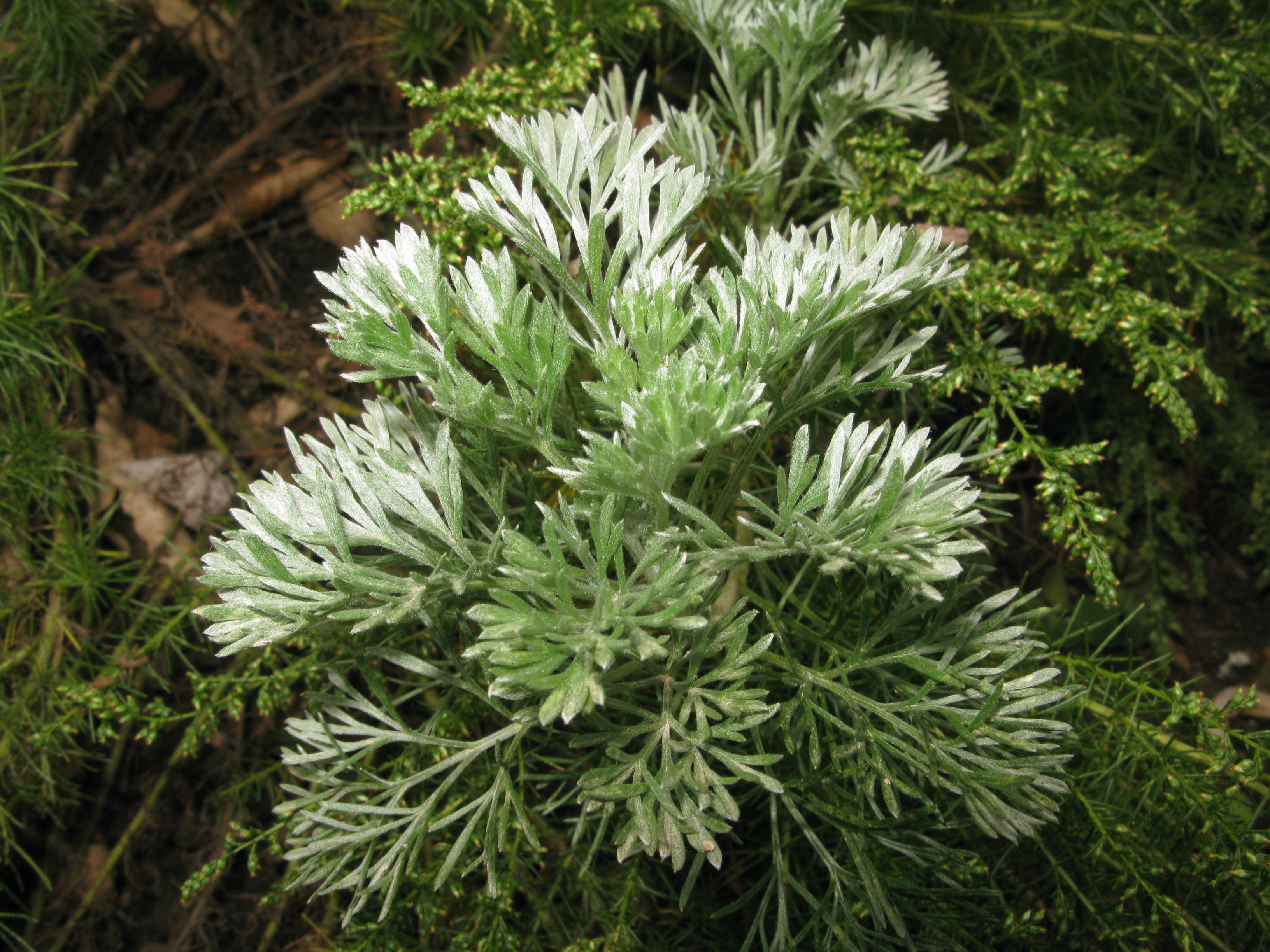
  • Development of a Natural Ointment Using Active Ingredient Content and Antibacterial Activity of Extracts from Ten Plants Including Artemisia capillaris
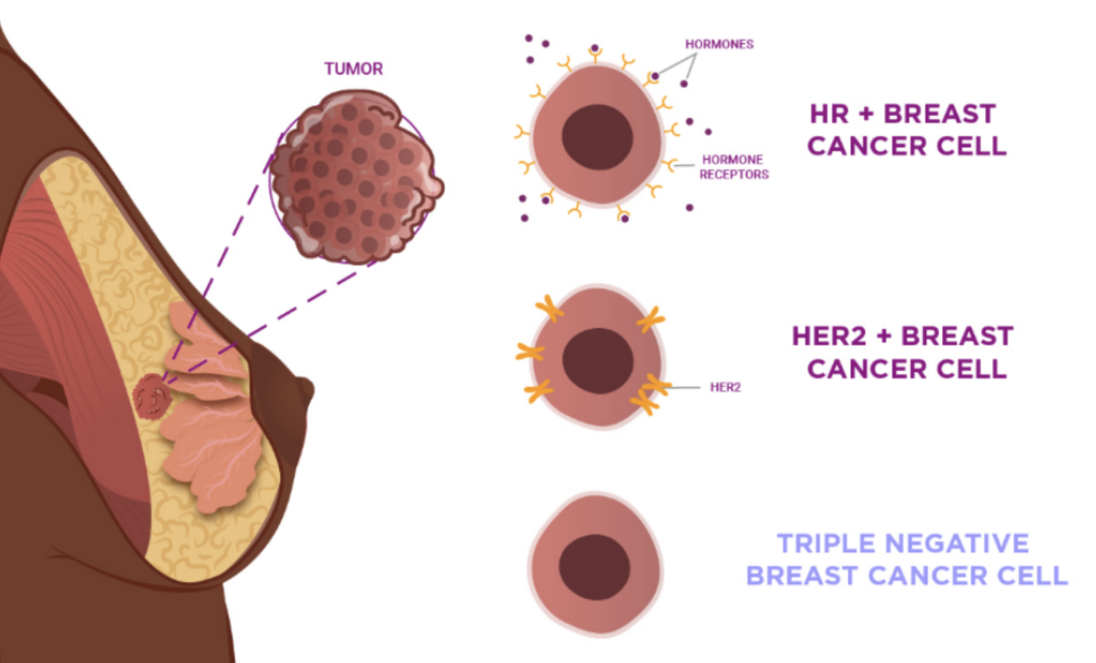
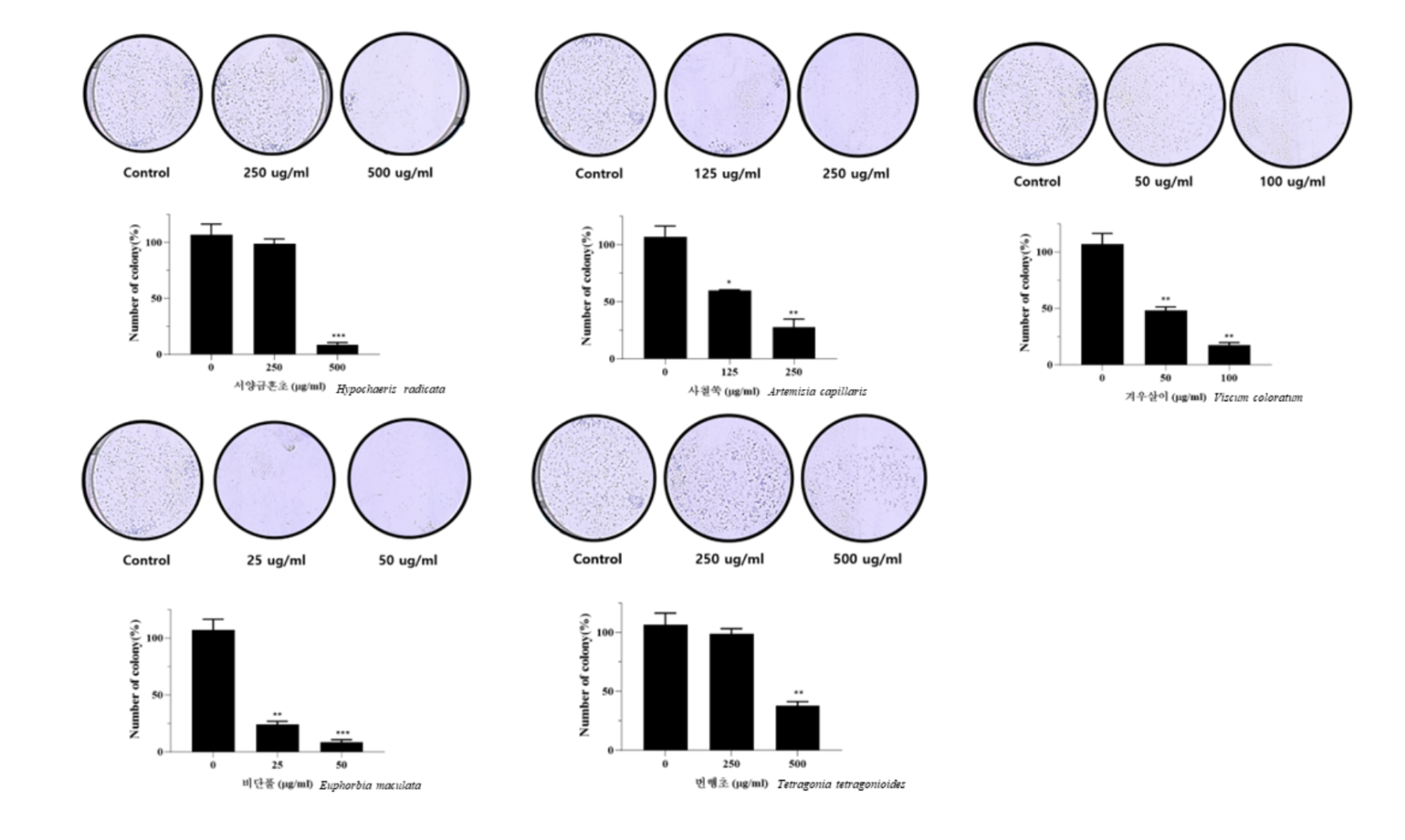
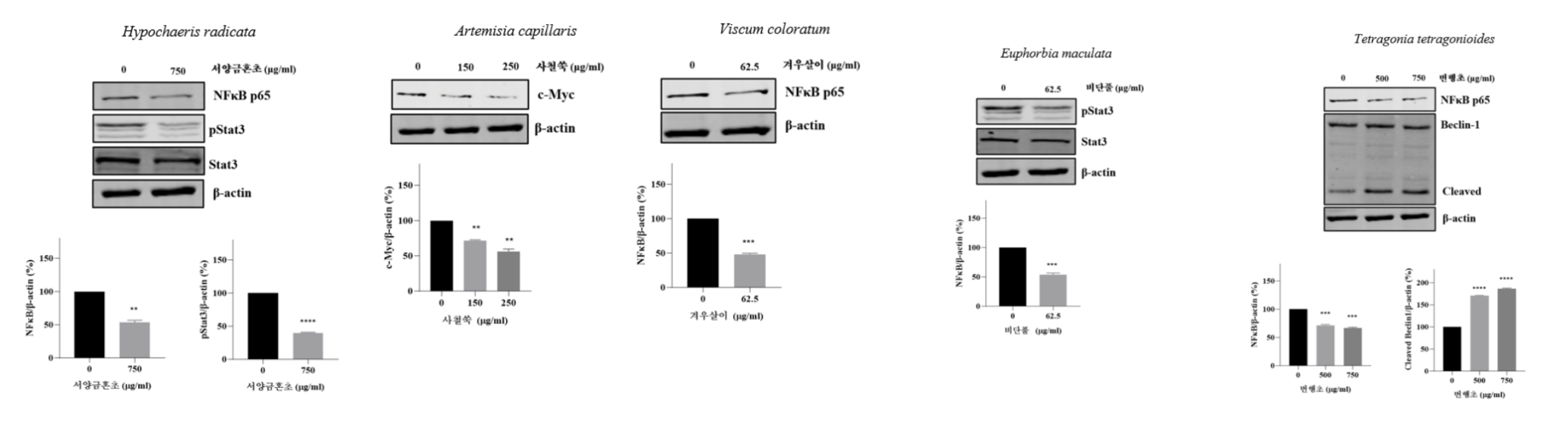
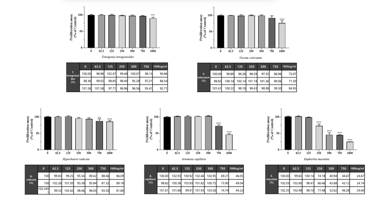
  • A Systematic Analysis of the Anticancer Activities of Medicinal Plant Extracts in Triple-Negative Breast Cancer (MDA-MB-231) cells
  • Analysis of antibacterial activity of acne bacteria found in Sasa quelpaertensis extract and efficacy as cosmetics

Future Goals

I want to pursue biology and continue developing innovative solutions to real-world health and environmental problems.

YeJin Ahn Lahye Yun

YeJin Ahn

St. Johnsbury Academy Jeju (SJA), Grade 11

Interests:

  • Cosmetics
  • Watching scientific videos (mostly about nature)

Research Projects:

  • Sasa Quelpaertensis-Based Feminine Hygiene Wash
  • Jeju Jori Tea for Diabetes Management

Future Goals

Later, I hope to attend medical school and become a doctor, where I can continue exploring my interest in health, especially women's health.
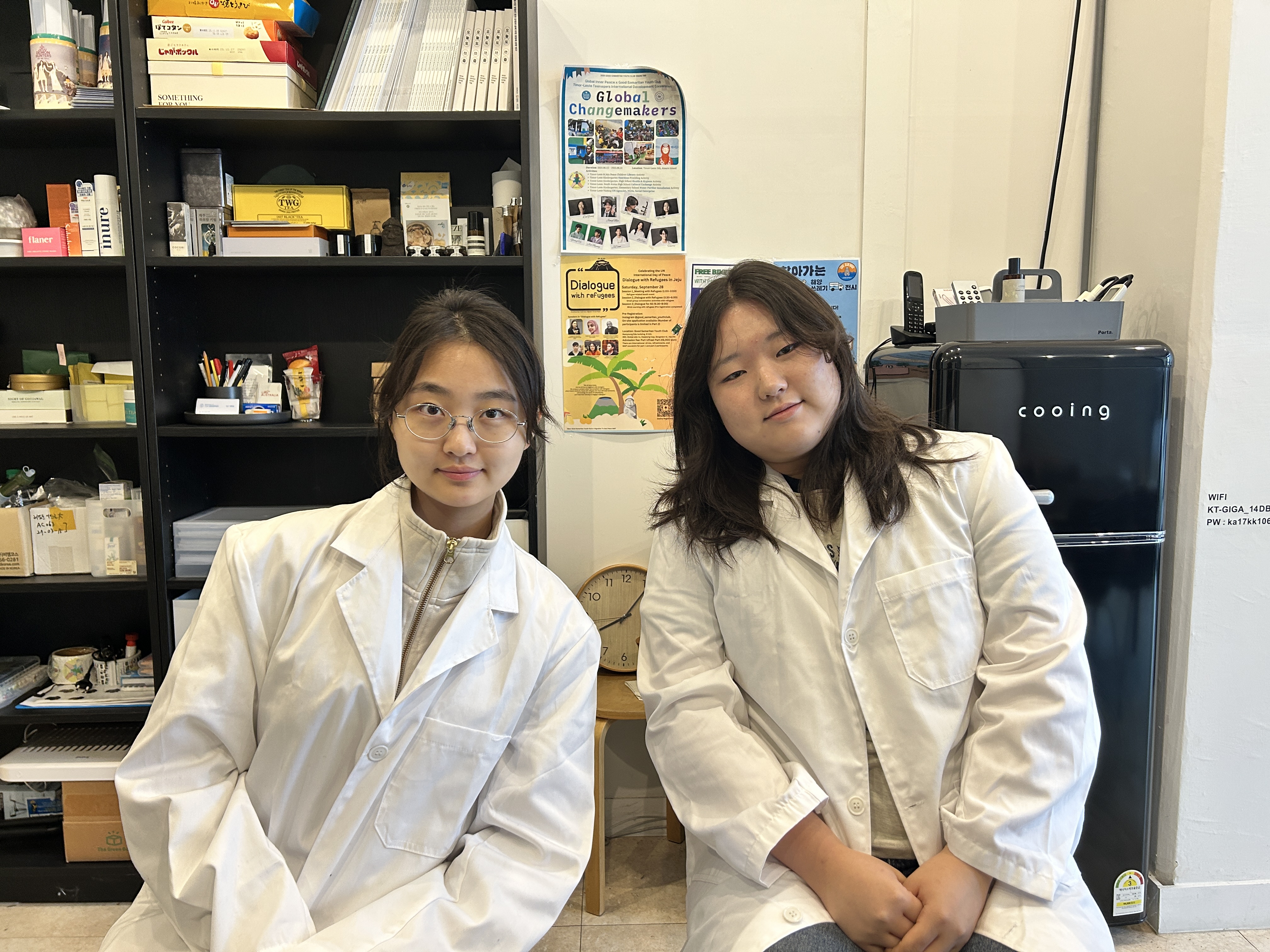
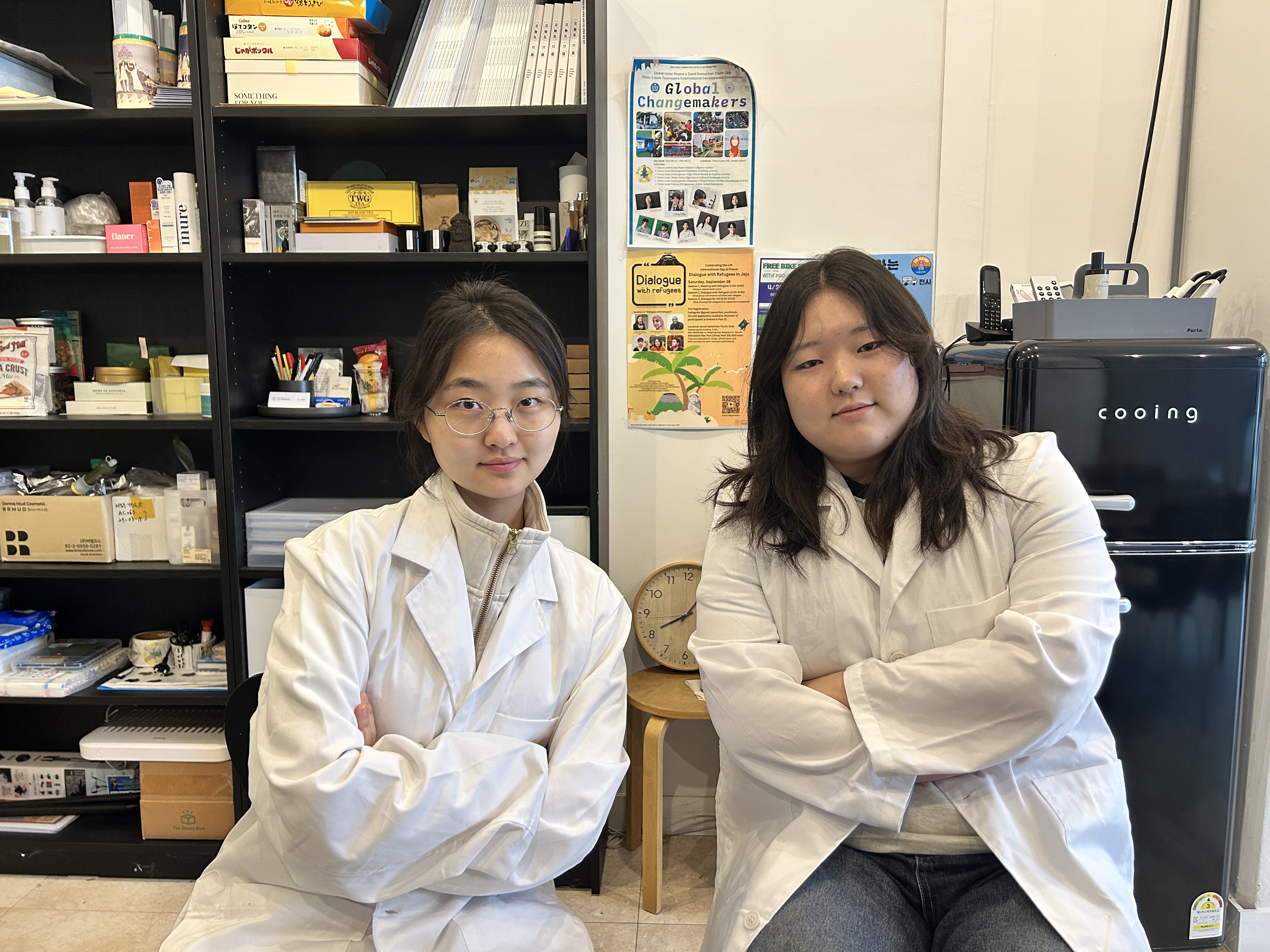
Research Team Research Team

Who are We?

We are a female-led research team from Jeju. We are 11th graders, Lahye Yun and Yejin Ahn, and we conduct research using a variety of plant resources that grow on Jeju Island.

Background

While we were continuously conducting research using Jeju plant resources, we witnessed a close friend's mother struggle with the side effects of radiation therapy. That experience led us to ask, "Is there a safer therapeutic candidate?"
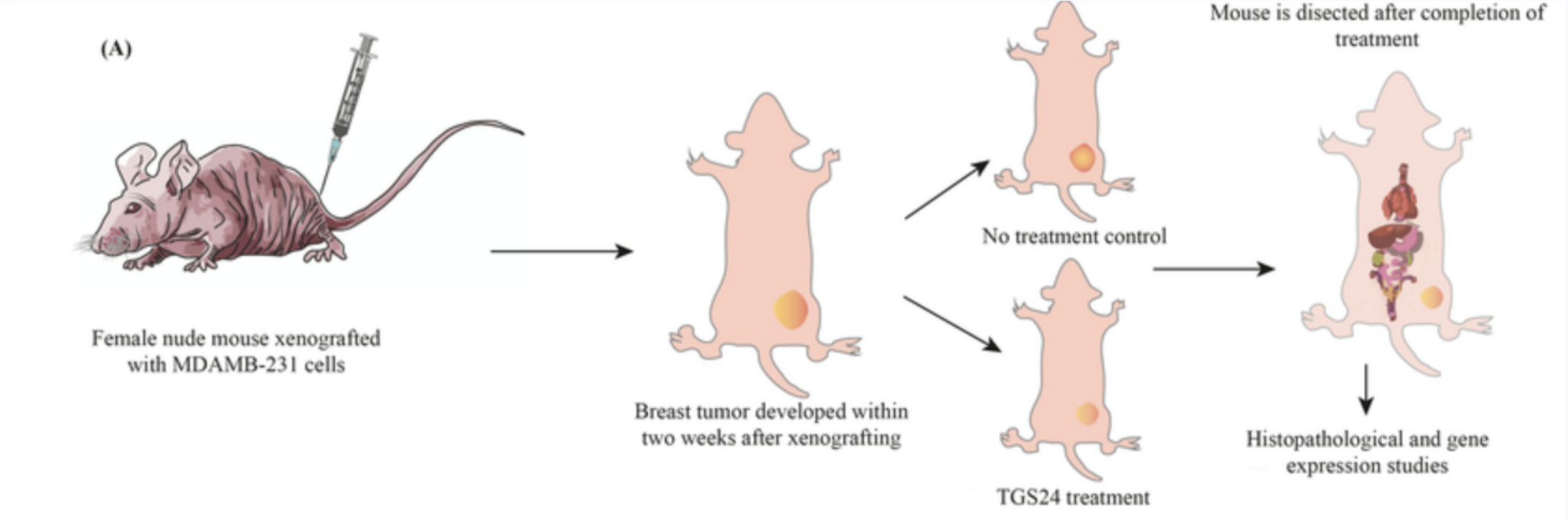
Goal

Using MDA-MB-231 cells, we aimed to explore biocompatible, plant-based therapeutic candidates from domestic plants that could complement the limitations of radiation therapy.

Research Background

Triple-Negative Breast Cancer

Triple-negative breast cancer lacks estrogen, progesterone, and HER2 receptors. Because these key receptors are absent, this does not respond to targeted or hormone-based treatments and is considered the most difficult form of breast cancer to treat.

In this study, we used the MDA-MB-231 cell line, which represents triple-negative breast cancer, an aggressive form that accounts for about 15-20% of cases.

Breast Duct Anatomy Cancer Cell Types

Limitations of Current Therapies

Treatment for triple-negative breast cancer mainly relies on chemotherapy and radiation therapy due to the limited effectiveness of targeted therapy. However, commonly used chemotherapeutic drugs cause serious side effects.

Cisplatin

Induces nephrotoxicity, along with damaging both cancer cells and normal cells.

Radiation Therapy

Significant side effects affecting overall patient quality of life and immune function.

Plant-Derived Therapeutic Potential

Selected Plant Species

Hypochaeris

Hypochaeris radicata

Artemisia

Artemisia capillaris

Viscum

Viscum coloratum

Euphorbia

Euphorbia maculata

Tetragonia

Tetragonia tetragonioides

Current applications of plant-derived compounds in cancer treatment highlight their significant therapeutic potential. Their lower toxicity and established safety profiles highlight the need for effective plant-derived treatments for triple-negative breast cancer.

Research Process

Plant Extraction Method

1

Collection

Collect plants from Jeju Island

2

Grinding

Grind into fine powder

3

Extraction

Ultrasound-assisted extraction

4

Freeze-Drying

Filtration, concentration, freeze-drying

Experimental Methods

1. MTT Cell Viability Assay

Confirm cytotoxicity and cell proliferation inhibitory effects

1
2

2. Wound Healing Assay

Evaluate cell migration inhibition

3. Colony Formation Assay

Long-term proliferation assessment

3
4

4. Apoptosis & Cell Cycle Analysis

Flow cytometry-based analysis

5. qPCR & Western Blot

mRNA and protein expression analysis

5

Experimental Methods

01

MTT Assay

  • Cell seeding: 1x10^4 cells/well
  • 96-well plates, 24h incubation
  • Extract treatment: 0-1000 ug/mL
  • MTT: 0.5 mg/mL, 2-4 hours
  • Measurement at 570 nm
02

Wound Healing

  • 6-well plates, confluent monolayer
  • Scratch with 200 uL pipette tip
  • 0.5% FBS assay medium
  • Monitoring: 0, 6, 12, 16 hours
03

Colony Formation

  • 6-well plates, 1,000 cells/well
  • 10-14 days culture
  • Acetic acid:methanol (3:1) fixation
  • Crystal violet (0.5%) staining
04

Apoptosis Assay

  • Annexin V/PI dual staining
  • BD Pharmingen Detection Kit
  • Flow cytometry analysis
  • Live, early/late apoptosis, necrotic
05

Cell Cycle

  • Cold 70% ethanol fixation
  • PI + RNase A staining
  • DNA-content histograms
  • G0/G1 and S/G2M analysis
06

qPCR

  • TRI-reagent RNA extraction
  • 260/280 purity check
  • SYBR Green, 40 cycles
  • GAPDH normalization
07

Western Blot Analysis

  • RIPA buffer protein extraction
  • BCA quantification
  • 15 ug protein, 10% SDS-PAGE
  • PVDF membrane transfer

Primary Targets:

NF-kB p65 p-STAT3 STAT3 c-Myc beclin-1

Results

MTT Cell Viability Assay

Before using the extracts in our experiments, we first needed to confirm cytotoxicity.

MTT Cell Viability Assay
Click to enlarge

We found that Hypochaeris and Tetragonia showed little cytotoxicity (overall), whereas Viscum, Artemisia, and Euphorbia showed cytotoxic effects at higher concentrations.

Wound Healing Assay

For the wound-healing assay, we cultured the cells in 6-well plates, created a scratch using a pipette tip, and then treated the cells with each extract.

Wound Healing Assay
Click to enlarge

In the control group, the wound area decreased over time. In contrast, when the extracts were applied, the wound area remained largely unchanged, indicating that cell migration and proliferation were inhibited.

Colony Formation Assay

In the long-term culture (colony formation) assay, we observed that all extracts clearly suppressed the ability of MDA-MB-231 cells to form colonies.

Colony Formation Assay
Click to enlarge

These results indicate that each extract has a strong antiproliferative effect, inhibiting the cells' ability to divide and maintain colonies in a concentration-dependent manner.

Western Blot Analysis

Western blot analysis supported the qPCR results at the protein level.

Western Blot Analysis
Click to enlarge

Western blot analysis supported these results. Hypochaeris reduced p65 and pSTAT3 expression, while Artemisia decreased c-Myc levels. Viscum showed a dual cell death-inducing pattern, with reduced p65 expression and activation of the RIPK1, RIPK3, and MLKL pathway, indicating involvement of multiple cell death mechanisms.

Cytotoxicity Evaluation

Evaluation of cytotoxicity across all plant extracts.

Cytotoxicity Evaluation
Click to enlarge

Evaluation of cytotoxicity revealed that Tetragonia tetragonoides and Hypochaeris radicata exhibited minimal toxicity even at high concentrations, while other extracts demonstrated dose-dependent effects, highlighting the importance of optimizing concentration ranges for safe therapeutic use.

Result Summary

Hypochaeris

Hypochaeris radicata

  • Inhibits wound healing and long-term colony formation
  • Significant results from an invasive species
  • Minimal toxicity even at high concentrations
  • Reduced p65 and pSTAT3 expression
Artemisia

Artemisia capillaris

  • Strongly inhibits cell migration and colony formation
  • Induces G2/M arrest
  • Decreased c-Myc levels
Tetragonia

Tetragonia tetragonioides

  • Shows little to no toxicity at low concentrations
  • Reduced NF-kB p65 expression
  • Increased Beclin-1 cleavage
Euphorbia

Euphorbia maculata

  • Inhibits cell migration and colony formation
  • Higher necrotic and dead populations
  • Decreased pSTAT3 while total STAT3 unchanged
Viscum

Viscum coloratum

  • Shows effects even at low concentrations
  • Dual cell death-inducing pattern
  • Reduced p65 expression and RIPK pathway activation

Key Findings

Because each experiment used different treatment concentrations, we cannot directly compare anticancer potency across plants under a standardized condition. Instead, we focused on whether each extract exhibited anticancer effects in MDA-MB-231 cells and on the mechanisms underlying those effects.

Most Promising Candidates

Tetragonia, Artemisia, and Hypochaeris show the greatest potential, given their bioactive compounds as well as their environmental and economic advantages. However, Viscum and Euphorbia should still be retained as potential candidates for further research.

Future Experiments

HPLC

HPLC & LC-MS

For next steps, we will identify active compounds using HPLC and LC-MS analyses to pinpoint the specific molecules driving the observed anticancer effects.

MPLC

MPLC

Isolate and test individual fractions through MPLC to determine which specific components or synergistic combinations are responsible for the results.

Mouse Model

Animal Studies

While not feasible at this stage, we plan to pursue in vivo validation in future university research, with the goal of ultimately developing a viable therapeutic product.